Spinal MRI; diagnosis the cause of low back pain and herniated disc
A spine MRI, or magnetic resonance imaging, uses powerful magnets, radio waves, and a computer to make very clear and detailed pictures of your spine. You may need this scan to check for spine problems such as Low back pain, Neck pain and Numbness, tingling, and weakness in your arms and legs. The MRI may scan your whole spine or just a part of it. Unlike X-rays and CT scans, it doesn’t use damaging radiation.
Anatomy of the spine
 The spinal column is made up of 33 vertebrae that are separated by spongy disks and classified into distinct areas.
- The cervical area consists of 7 vertebrae in the neck.
- The thoracic area consists of 12 vertebrae in the chest area.
- The lumbar area consists of 5 vertebrae in the lower back area.
- The sacrum has 5 small, fused vertebrae.
- The 4 coccygeal vertebrae fuse to form 1 bone, called the coccyx or tailbone.
The spinal cord, a major part of the central nervous system, is located in the vertebral canal and reaches from the base of the skull to the upper part of the lower back. The spinal cord is surrounded by the bones of the spine and a sac containing cerebrospinal fluid. The spinal cord carries sensory and movement signals to and from the brain, and controls many reflexes.
why is MRI done?

MR imaging is performed to:
- assess spinal anatomy and alignment.
- detect congenital anomalies of vertebrae or the spinal cord.
- detect bone, disc, ligament or spinal cord injury after spine trauma.
- assess intervertebral disk disease such as degenerated, bulging or herniated and intervertebral joint disease, both frequent causes of severe lower back pain and sciatica (back pain radiating into lower leg).
- explore other possible causes of back pain (compression fracture or bone swelling, such as edema).
- assess compression of spinal cord and nerves.
- assess inflammation of the spinal cord or nerves.
- assess infection involving the spine, disks and spinal contents including spinal cord or its coverings.
- assess tumors that arise from or have spread to the vertebrae, spinal cord, nerves or the surrounding soft tissues.
- help plan spinal surgical procedures, such as decompression of a pinched nerve, spinal fusion, or the injection of steroids to relieve spinal pain. Such injections are usually performed under CT guidance.
- monitor changes in the spine after an operation, such as scarring or infection.
Pre-spinal MRI Care
EAT/DRINK
You may eat, drink and take medications as usual.
CLOTHING
You must completely change into a patient gown and lock up all personal belongings. A locker will be provided for you to use. Please remove all piercings and leave all jewelry and valuables at home.
ALLERG
If you have had an allergic reaction to contrast that required medical treatment, contact your ordering physician to obtain the recommended prescription. You will likely take this by mouth 24, 12 and two hours prior to examination.
ANTI-ANXIETY MEDICATION
If you require anti-anxiety medication due to claustrophobia, contact your ordering physician for a prescription. Please note that you will need some else to drive you home.
The presence of metals in the body
If you have metal within your body that was not disclosed prior to your appointment, your study may be delayed, rescheduled or cancelled upon your arrival until further information can be obtained. Based on your medical condition, your doctor may require other specific preparation. When you call to make an appointment, it is extremely important that you inform if any of the following apply to you:
- You have a pacemaker or have had heart valves replaced
- You have any type of implantable pump, such as an insulin pump
- You have metal plates, pins, metal implants, surgical staples or aneurysm clips
- You are pregnant or think you might be pregnant
- You have any body piercing
- You are wearing a medication patch
- You have permanent eye liner or tattoos
- You have ever had a bullet wound
- You have ever worked with metal (for example, a metal grinder or welder)
- You have metallic fragments anywhere in the body
- You are not able to lie down for 30 to 60 minutes.
What's the Equipment Like?
An MRI machine is a long, narrow tube with both ends open. A magnet surrounds the tube. You lie on a table that slides into the tube. Some MRI machines have much larger openings or are open on the sides so you don’t have to slide into a tube. They may be a good choice if you are overweight or fear tight spaces. Your doctor will decide which MRI machine will work best for you.
How to do Lumbar MRI-min?
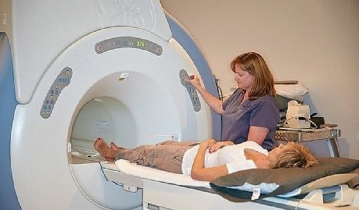
Before some MRIs, you might need a dye injected into a vein in your arm or hand. It helps the doctor more clearly see any infection, tumor, or disk problem in your spine. The dye often used in MRIs is called gadolinium. You might feel flush or cold for a few moments afterward. It can also leave a salty or metal taste in your mouth. You’ll lie on the table that slides into the MRI machine. Straps may be used to help keep you in the right position during the test. A radiologist and technologist will be at a computer, outside of the room. They can see, hear, and talk to you the whole time. Sometimes your family or friend can stay in the room with you. Often an MRI exam includes a number of runs, or sequences. Each run can last from a few seconds to several minutes long. You have to stay very still during each one. The MRI machine creates a strong magnetic field around you. A computer takes the signals from the MRI and uses them to make a series of pictures. Each picture shows a thin slice of your body. You won’t feel any pain during the test. But you may feel warmth in the area of your spine being scanned. You’ll also hear a loud tapping or thumping when the image is being recorded. Earplugs or headsets can help block out the noise if it bothers you. You can even listen to music. MRI scans can take from 30 minutes to an hour or more, depending on how much of your spine is being scanned. After a spine MRI, you can go back to your normal activities right away. But if you needed medicine to relax before the test, you’ll need to wait until it wears off.
Post-spinal MRI Care

You should move slowly when getting up from the scanner table to avoid any dizziness or lightheadedness from lying flat for the length of the procedure. If any sedatives were taken for the procedure, you may be required to rest until the sedatives have worn off. You will also need to avoid driving. If contrast dye is used during your procedure, you may be monitored for a period of time for any side effects or reactions to the contrast dye, such as itching, swelling, rash, or difficulty breathing. If you notice any pain, redness, and/or swelling at the IV site after you return home following your procedure, you should notify your doctor as this could indicate an infection or other type of reaction. Otherwise, there is no special type of care required after a MRI scan of the spine. You may resume your usual diet and activities, unless your doctor advises you differently. Your doctor may give you additional or alternate instructions after the procedure, depending on your particular situation.
What are the limitations of MRI of the Spine?
-
High-quality images are assured only if you are able to remain perfectly still and follow breath-holding instructions while the images are being recorded. If you are anxious, confused or in severe pain, you may find it difficult to lie still during imaging.
- A person who is very large may not fit into the opening of certain types of MRI machines.
- The presence of an implant or other metallic object sometimes makes it difficult to obtain clear images due to streak artifacts from the metallic objects. Patient movement can have the same effect.
- A very irregular heartbeat may affect the quality of images obtained using techniques that time the imaging based on the electrical activity of the heart, such as electrocardiography (EKG).
-
MRI generally is not recommended for patients who have been acutely injured; however, this decision is based on clinical judgment. This is because traction devices and many types of life support equipment may distort the MR images and as a result, must be kept away from the area to be imaged. Furthermore, the examination takes longer than other imaging modalities (typically x-ray and CT) and the results may not be immediately available, as is often necessary in trauma situations.
- Although there is no reason to believe that magnetic resonance imaging harms the fetus, pregnant women usually are advised not to have an MRI exam during the first trimester unless medically necessary.
- MRI typically costs more and may take more time to perform than other imaging modalities.
- In some patients, vertebral fractures may be better detected by CT scan.
Risks and complications of MRI
- The MRI examination poses almost no risk to the average patient when appropriate safety guidelines are followed.
- If sedation is used, there are risks of excessive sedation. However, the technologist or nurse will monitor your vital signs to minimize this risk.
- Although the strong magnetic field is not harmful in itself, implanted medical devices that contain metal may malfunction or cause problems during an MRI exam.
- Nephrogenic systemic fibrosis is currently a recognized, but rare, complication of MRI believed to be caused by the injection of high doses of gadolinium-based contrast material in patients with very poor kidney function. Careful assessment of kidney function before considering a contrast injection minimizes the risk of this very rare complication.
- There is a very slight risk of an allergic reaction if contrast material is injected. Such reactions are usually mild and easily controlled by medication. If you experience allergic symptoms, a doctor will be available for immediate assistance.